 Wake Forest Institute for Regenerative Medicine scientists (WFIRM) have developed a method to bioprint a type of cartilage that could someday help restore knee function damaged by arthritis or injury.
This cartilage, known as fibrocartilage, helps connect tendons or ligaments or bones and is primarily found in the meniscus in the knee. The meniscus is the tough, rubbery cartilage that acts as a shock absorber in the knee joint. Degeneration of the meniscus tissue affects millions of patients, and arthroscopic partial meniscectomy is one of the most common orthopedic operations performed. Besides surgery, there is a lack of available treatment options.
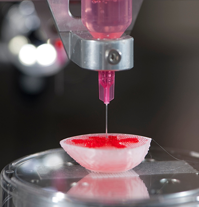 In this latest proof-of-concept strategy, the scientists were able to 3D bioprint a hybrid tissue construct for cartilage regeneration by printing two specialized bioinks – hydrogels that contain the cells – together to create a new formulation that provides a cell-friendly microenvironment and structural integrity. This work is done with the Integrated Tissue and Organ Printing System, a 3D bioprinter that was developed by WFIRM researchers over a 14-year period. The system deposits both biodegradable, plastic-like materials to form the tissue “shape” and bioinks that contain the cells to build new tissues and organs.
“In this study, we have been able to produce a highly elastic hybrid construct for advanced fibrocartilaginous regeneration,” said Sang Jin Lee, PhD, associate professor at WFIRM and author of the paper published in fall 2020 by Chemistry of Materials journal. “The results demonstrate that this bioprinted construct offers a versatile and promising alternative for the production of this type of tissue.”